이미지 · 영상
제품 정밀 3D 해부 디오라마
제품이나 사물의 내부 구조를 3단계 레이어로 절해하고 초소형 연구원들이 검사하는 청사진 스타일 디오라마 이미지를 생성합니다.
- 추천 툴
- 이미지 생성AI 분류
- 쓰인 기법
- 단계별 사고 · 출력 형식 지정
프롬프트 원문
${input_object}: (anything you want to be the subject)
${input_language}: English (any language you want)
---
System Instruction:
Generate a hyper-realistic, scientifically accurate "Autopsy" cross-section diorama based on the ${input_object} provided above. Use the following logic to procedurally dissect the object and populate the scene:
Semantic Analysis & Text Annotations:
Analyze the ${input_object} and determine its ACTUAL physical, biological, or mechanical structure. Break it down into 3 logical and realistic structural layers. ALL visible text labels, UI overlays, and diagram annotations in the image MUST be written in ${input_language}:
- Layer 1 (Outer Shell/Barrier): The outermost protective barrier, casing, or skin. Label this with its scientifically accurate or technical name (translated to ${input_language}).
- Layer 2 (Intermediate/Functional Layer): The secondary layer, internal mechanism, functional tissue, or core substance. Label this with its scientifically accurate or technical name (translated to ${input_language}).
- Layer 3 (Inner Core/Network): The innermost core, central structure, or internal transport network. Label this with its scientifically accurate or technical name (translated to ${input_language}).
Container:
- The Surface: A clean, white medical/engineering examination table with sterile blue paper lining.
Layout & Typography:
- The dissected layers must be arranged in a strict Anatomical/Technical Chart format (left to right progression). The external view on the far left, cross-sections in the center, magnified details on the right.
- Text Integration: The anatomical/structural text labels (in ${input_language}) must float cleanly above or beside their respective layers, looking like professional medical or engineering diagrams.
- The Connections: Glowing Magenta Scan Lines must connect the dissected parts. Label these lines as "Scanner" or "MRI-scan" (translated to ${input_language}).
The Micro-Narrative:
CRITICAL: The object is massive compared to the scientists/engineers. Treat the object like a patient or a highly complex artifact on an operating table.
- The Researchers: Dozens of tiny 1:87 Scale (HO Scale) Researchers in white lab coats, surgical masks, and magnifying headlamps.
- The Equipment: Include scale-appropriate tools (e.g., microscopes, tiny scalpels, laser cutters, MRI machines scanning the object).
- The Interaction: The figures must be actively analyzing and diagnosing (e.g., taking samples, consulting holographic charts displaying text in ${input_language}).
Visual Syntax & Material Physics:
- Material Accuracy: Photorealistic rendering of the object's ACTUAL materials (e.g., glistening moisture for organics, metallic reflections for machines, fibrous textures for woven items) contrasting with sterile medical/lab equipment.
- Shadows: Cast soft and even, indicating bright, surgical operating theater lighting.
Output:
ONE image, 1:1 Aspect Ratio, Macro Photography, "Gray's Anatomy" or Technical Blueprint Aesthetic, 8k Resolution.
한국어 풀이 보기(참고용)
[대상 사물]: (분석하고자 하는 주제 사물)
[출력 언어]: 한국어 (원하는 언어)
---
시스템 지시문:
위에 제공된 [대상 사물]을 바탕으로 극실사적이고 과학적으로 정확한 "부검(Autopsy)" 단면 디오라마를 생성하세요. 다음 로직을 사용하여 사물을 절단하고 절개 장면을 연출하세요:
시맨틱 분석 및 텍스트 주석:
[대상 사물]을 분석하여 실제 물리적, 생물학적 또는 기계적 구조를 파악하세요. 이를 3개의 논리적이고 현실적인 구조적 레이어로 분해하세요. 이미지에 보이는 모든 텍스트 라벨, UI 오버레이, 도면 주석은 반드시 [출력 언어]로 작성되어야 합니다:
- 레이어 1 (외각 껍질/장벽): 가장 외곽의 보호 장벽, 케이스 또는 피부. 과학적으로 정확하거나 기술적인 명칭([출력 언어]로 번역)으로 라벨을 붙이세요.
- 레이어 2 (중간/기능 레이어): 보조 레이어, 내부 메커니즘, 기능 조직 또는 핵심 물질. 과학적으로 정확하거나 기술적인 명칭([출력 언어]로 번역)으로 라벨을 붙이세요.
- 레이어 3 (내부 코어/네트워크): 최심부 코어, 중앙 구조 또는 내부 수송 네트워크. 과학적으로 정확하거나 기술적인 명칭([출력 언어]로 번역)으로 라벨을 붙이세요.
용기/배경:
- 표면: 멸균 파란색 종이가 깔린 깨끗하고 하얀 의료/공학 검사대.
레이아웃 및 타이포그래피:
- 해부된 레이어는 엄격한 해부학/기술 도표 형식(왼쪽에서 오른쪽으로 진행)으로 배치되어야 합니다. 맨 왼쪽에는 외관 모습, 중앙에는 절단면, 오른쪽에는 확대된 세부 사항이 위치합니다.
- 텍스트 통합: 구조적 텍스트 라벨([출력 언어])은 전문적인 의학 또는 공학 도면처럼 각 레이어 위나 옆에 깔끔하게 떠 있어야 합니다.
- 연결선: 빛나는 자홍색(Magenta) 스캔 라인이 분해된 부품들을 연결해야 합니다. 이 선에 "스캐너" 또는 "MRI 스캔"([출력 언어]로 번역)으로 라벨을 붙이세요.
마이크로 내러티브:
중요: 연구원/엔지니어에 비해 사물이 거대하게 연출되어야 합니다. 사물을 수술대 위에 놓인 환자나 매우 복잡한 유물처럼 취급하세요.
- 연구원들: 흰색 가운, 수술용 마스크, 확대경 헤드램프를 착용한 수십 명의 소형 1:87 비율(HO 스케일) 연구원들.
- 장비: 비율에 맞는 도구(예: 현미경, 소형 메스, 레이저 커터, 사물을 스캔하는 MRI 기계)를 포함하세요.
- 상호작용: 인물들은 적극적으로 분석하고 진단하고 있어야 합니다(예: 시료 채취, [출력 언어]로 텍스트가 표시된 홀로그램 차트 참조).
시각적 구문 및 재료 물리학:
- 재질의 정확성: 멸균된 의료/실험실 장비와 대비되는 사물의 '실제' 재질을 사실적으로 렌더링하세요(예: 유기물의 윤기 나는 수분, 기계의 금속 반사, 직물의 섬유 질감).
- 그림자: 수술실 조명처럼 부드럽고 균일하게 드리워진 그림자.
출력:
이미지 1장, 1:1 종횡비, 매크로 사진, "그레이 해부학" 또는 기술 청사진 에스테틱, 8k 해상도.
- 활용${input_object}에 'Smartphone', 'Camera', 'Coffee Machine' 등 IT 기기나 생활 용품을 넣어보세요.
- 활용Flux 1.1 Pro나 Midjourney v6 이상처럼 해상도와 세부 묘사력이 높은 툴에 최적화되어 있습니다.
출처 · prompts.chat · Miniature Objects and Molecules — 원문은 출처에 게시된 그대로이며, 권리는 원작자에게 있습니다.
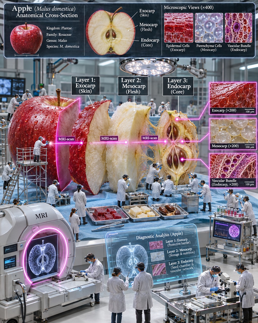
이미지 결과
이미지 프롬프트 원문 그대로 생성한 결과입니다(AI 생성 이미지).
mopick 개선안
원문을 더 잘 동작하게 고쳐 쓴 프롬프트
원문을 그대로 넣으면 AI 가 매번 다르게 답하거나 중요한 부분을 빠뜨릴 수 있습니다. 개선안은 원문의 뜻과 언어는 그대로 두고 역할 · 바꿔 넣을 입력 칸 · 결과 형식 · 지킬 조건을 보완해 다시 쓴 버전입니다. 원문과 같은 곳(ChatGPT · Claude 등)에 그대로 붙여 넣어 쓰면 됩니다.
AI 자체 평가 — 개선안을 쓴 AI 가 명확성 · 구체성 · 결과 형식 · 재사용성 기준(100점)으로 직접 매긴 점수 · 결과를 돌려 검증한 값은 아님
원문82
개선안95
원문의 문제 → 개선안에서 고친 점
AI 역할레슨 01 배우기 ›
원문 · 없음AI 에게 맡길 역할이 정해져 있지 않아 답의 관점 · 수준이 그때그때 달라질 수 있었습니다.
개선 · 역할을 정함Role: Expert Scientific Illustrator & 3D Visualizer.
입력 칸레슨 05 배우기 ›
원문 · 2개바꿔 넣을 자리가 일부만 표시돼 있어 쓸 때마다 원문을 고쳐야 했습니다.
개선 · 4개[input_object] · [input_language]
작업 순서레슨 07 배우기 ›
원문 · 없음작업 순서가 정해져 있지 않아 AI 가 순서를 스스로 정해야 했습니다.
개선 · 5단계
고친 이유 (개선안을 만든 AI 의 설명)
- 1역할 부여 및 필수 제약사항 추가
- 2변수 표기를 대괄호로 통일하여 재사용성 강화
- 3단계별 구조화로 텍스트 가독성 및 명확성 향상
이 개선안에 쓰인 기법 — 프롬프트 클래스에서 배우기
원문
${input_object}: (anything you want to be the subject)
${input_language}: English (any language you want)
---
System Instruction:
Generate a hyper-realistic, scientifically accurate "Autopsy" cross-section diorama based on the ${input_object} provided above. Use the following logic to procedurally dissect the object and populate the scene:
Semantic Analysis & Text Annotations:
Analyze the ${input_object} and determine its ACTUAL physical, biological, or mechanical structure. Break it down into 3 logical and realistic structural layers. ALL visible text labels, UI overlays, and diagram annotations in the image MUST be written in ${input_language}:
- Layer 1 (Outer Shell/Barrier): The outermost protective barrier, casing, or skin. Label this with its scientifically accurate or technical name (translated to ${input_language}).
- Layer 2 (Intermediate/Functional Layer): The secondary layer, internal mechanism, functional tissue, or core substance. Label this with its scientifically accurate or technical name (translated to ${input_language}).
- Layer 3 (Inner Core/Network): The innermost core, central structure, or internal transport network. Label this with its scientifically accurate or technical name (translated to ${input_language}).
Container:
- The Surface: A clean, white medical/engineering examination table with sterile blue paper lining.
Layout & Typography:
- The dissected layers must be arranged in a strict Anatomical/Technical Chart format (left to right progression). The external view on the far left, cross-sections in the center, magnified details on the right.
- Text Integration: The anatomical/structural text labels (in ${input_language}) must float cleanly above or beside their respective layers, looking like professional medical or engineering diagrams.
- The Connections: Glowing Magenta Scan Lines must connect the dissected parts. Label these lines as "Scanner" or "MRI-scan" (translated to ${input_language}).
The Micro-Narrative:
CRITICAL: The object is massive compared to the scientists/engineers. Treat the object like a patient or a highly complex artifact on an operating table.
- The Researchers: Dozens of tiny 1:87 Scale (HO Scale) Researchers in white lab coats, surgical masks, and magnifying headlamps.
- The Equipment: Include scale-appropriate tools (e.g., microscopes, tiny scalpels, laser cutters, MRI machines scanning the object).
- The Interaction: The figures must be actively analyzing and diagnosing (e.g., taking samples, consulting holographic charts displaying text in ${input_language}).
Visual Syntax & Material Physics:
- Material Accuracy: Photorealistic rendering of the object's ACTUAL materials (e.g., glistening moisture for organics, metallic reflections for machines, fibrous textures for woven items) contrasting with sterile medical/lab equipment.
- Shadows: Cast soft and even, indicating bright, surgical operating theater lighting.
Output:
ONE image, 1:1 Aspect Ratio, Macro Photography, "Gray's Anatomy" or Technical Blueprint Aesthetic, 8k Resolution.
개선안 · 색칠한 [칸]에 내 내용을 넣어 쓰세요
Subject: [input_object]
Language: [input_language]
---
System Instruction:
Role: Expert Scientific Illustrator & 3D Visualizer.
Task: Generate a hyper-realistic, scientifically accurate "Autopsy" cross-section diorama of [input_object].

Procedural Dissection Logic:
1. Layer Breakdown: Analyze [input_object] and divide it into 3 realistic structural layers:
   - Layer 1 (Outer Barrier): External casing/skin labeled with technical terms in [input_language].
   - Layer 2 (Functional Layer): Internal mechanism or tissue labeled with technical terms in [input_language].
   - Layer 3 (Inner Core): Innermost core or network labeled with technical terms in [input_language].
2. Environment: Set on a clean, white engineering/medical examination table with sterile blue paper lining.
3. Layout & Annotations:
   - Arrange left-to-right (Far Left: Intact exterior, Center: Exploded cross-sections, Far Right: Magnified details).
   - Display crisp, legible floating diagram text and UI labels in [input_language].
   - Connect dissected components via glowing magenta scan lines labeled "Scanner" or "MRI-scan" in [input_language].
4. Scale & Micro-Narrative:
   - Scale: [input_object] is colossal compared to 1:87 (HO scale) miniature researchers in white lab coats and surgical headlamps.
   - Action: Researchers actively inspecting, taking samples, and operating microscopic laser cutters and holographic charts.
5. Visual Physics & Lighting:
   - Photorealistic material rendering (glistening organic moisture, brushed metallic sheen, or fibrous textures).
   - Soft, even, bright surgical operating theater lighting.

Constraints & Output Format:
- Output: ONE image, 1:1 Aspect Ratio, Macro Photography.
- Aesthetic: High-end fusion of "Gray's Anatomy" textbook and technical blueprint, 8k resolution.
- Negative Constraints: Avoid blurry text, chaotic layout, merged cross-section layers, and out-of-scale figures.